
CBCT Reporting
What is included in a CBCT Report?
At Oral Radiologists, LLC, we put extensive time and effort into our reports, which are both comprehensive and detailed. We scrutinize every anatomical structure captured within the entire volume of a Cone Beam CT to rule out any type of pathologic or infectious process, within or outside of the oral cavity.
Within the oral cavity, we frequently unearth previously undetected caries, periapical inflammatory lesions, non-carious cervical lesions, periodontal disease, and other pathologic and inflammatory diseases. As a result of our reports, it is common that an immediate need for further dental treatment is discovered.
It is also common to encounter various findings outside the oral cavity which require follow-up by a medical provider. These include arterial calcifications, sinus diseases, skull base lesions, neck and TMJ arthritis, and any variety of cysts, tumors or infections in any of these anatomical structures.
What is the difference between a Standard and Detailed CBCT Report?
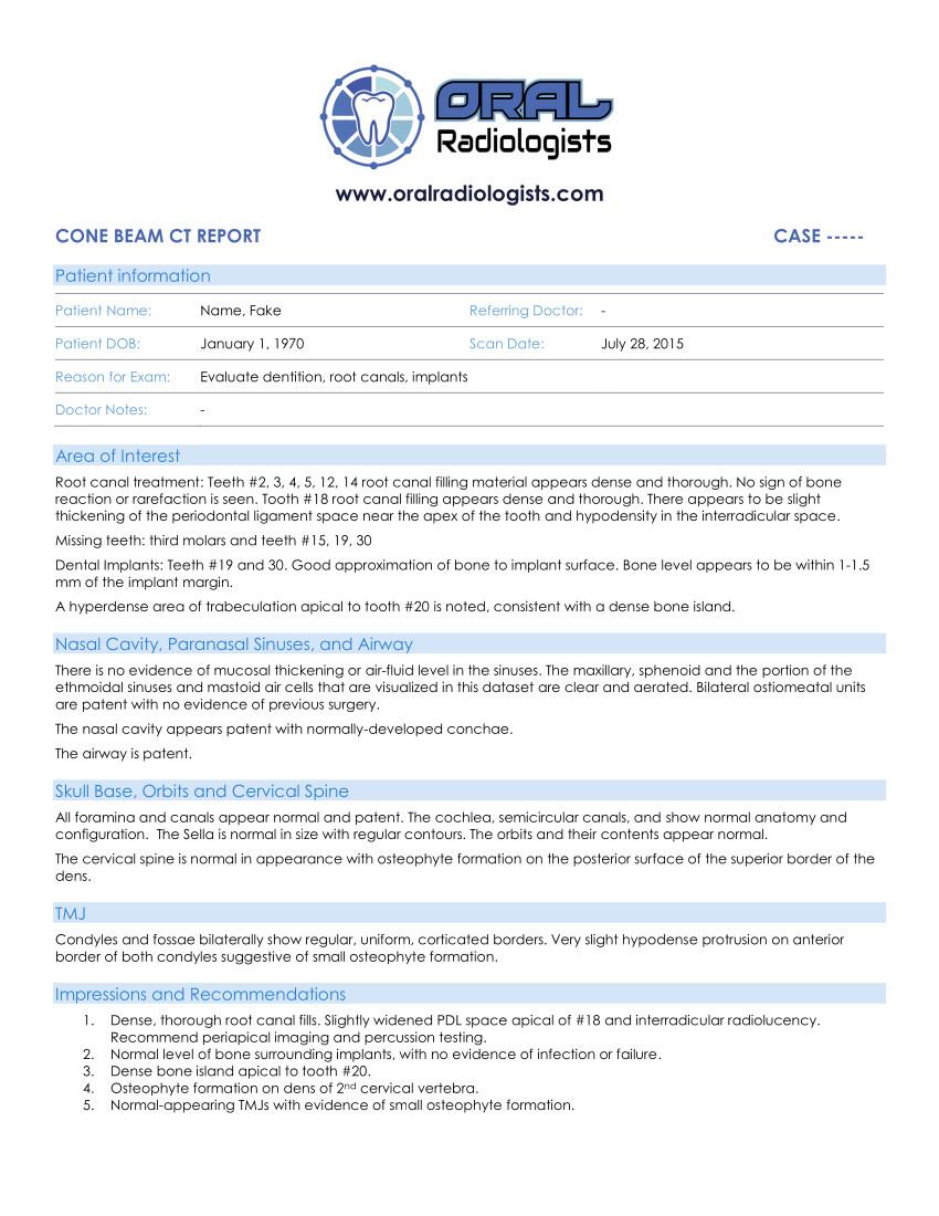
Standard CBCT Report
Both types of reports include thorough investigation of all structures found within a volume. A standard report will have a more basic description of the findings, perhaps a bullet point list of findings, without detailed description of the findings or its implications. It will still include an Impressions and Recommendations section, which delineates any clinically relevant findings and recommendations as appropriate. This section will include anything inside or outside of the oral cavity that will require further investigation, treatment, or referral to a medical provider.
A Standard Report will typically include images of a panoramic reformat of the oral cavity and up to 2-3 images of other pertinent findings.
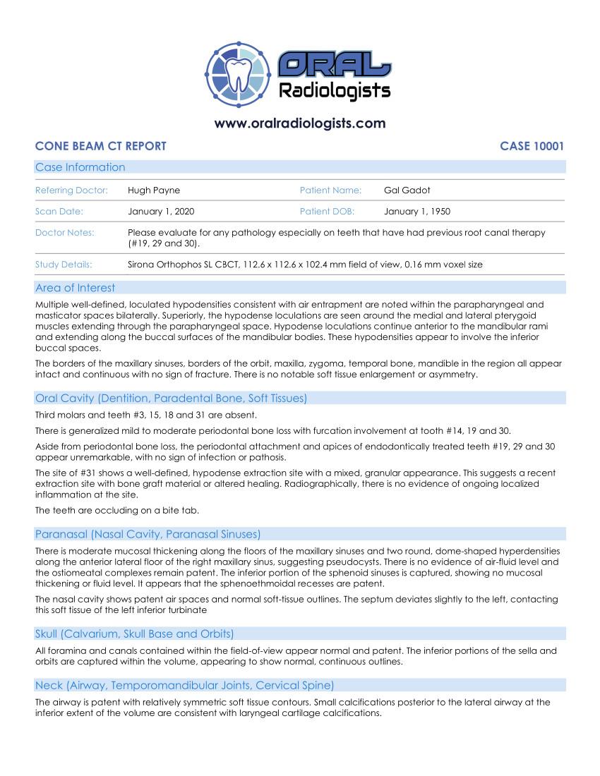
Detailed CBCT Report
A detailed report includes detailed description of all findings and is divided by anatomical category (Oral, Paranasal, Skull, Neck). This includes detailed description of different parts of an entity and its effect on other structures, and may include implications of features of an entity on potential diagnosis.
A detailed report will also include more images (as feasible) compared with the standard report. Images typically include a panoramic reformat of the oral cavity, oral airway analysis, TMJ view (if included in the volume), and images of several other important or incidental findings.
Like a standard report, the Detailed Report will include an Impressions and Recommendations section, delineating any clinically relevant findings and recommendations. It may also include the thought process behind why different diagnoses were considered when constructing a differential diagnosis.
If a report will be shared with a patient, another provider, or if you are interested in using the report as a tool for your own education and growth, it is recommended to request a Detailed CBCT Report. If there is a known pathologic or inflammatory condition you would like detail on, it is also recommended to request a Detailed CBCT Report.
See examples of a cone beam CT reports here....
Standard CBCT Radiology Report Example (with multiple intraoral findings and arterial calcifications)
Detailed CBCT Radiology Report Example (with subcutaneous emphysema and minimal intraoral findings)
For Instructions on submitting a case, CLICK HERE